

Utilizing cutting-edge dual-energy cone-beam X-ray imaging technology, the imaging mode can be seamlessly switched between single energy and dual energy. In single energy mode (DR mode), it facilitates rapid imaging and two-dimensional measurements in seconds for experimental animals. In dual energy mode (DXA mode), it further provides quantitative parameter information such as density and body composition.
The adoption of large-sized flat panel detectors has effectively increased the imaging area, enabling high-throughput imaging of multiple samples/mice;
Equipped with white LEDs and HD cameras, it precisely marks the animal placement area, enabling real-time monitoring and clear photography;
High-quality 304 stainless steel inner liner, easy to disinfect and clean.
Integrating image acquisition, data management, parameter analysis, and reporting functions;
It can also calculate parameters such as bone and body composition for the whole body and multiple ROIs, which can be used for relevant quantitative research;
The software interface is simple and clean, and it allows for the simultaneous display of X-ray images (including low-energy, high-energy, skeletal, and fat/lean meat images), optical images, and fused images in separate windows during image browsing. It supports the fusion of any number of X-ray and optical images.
.jpg)
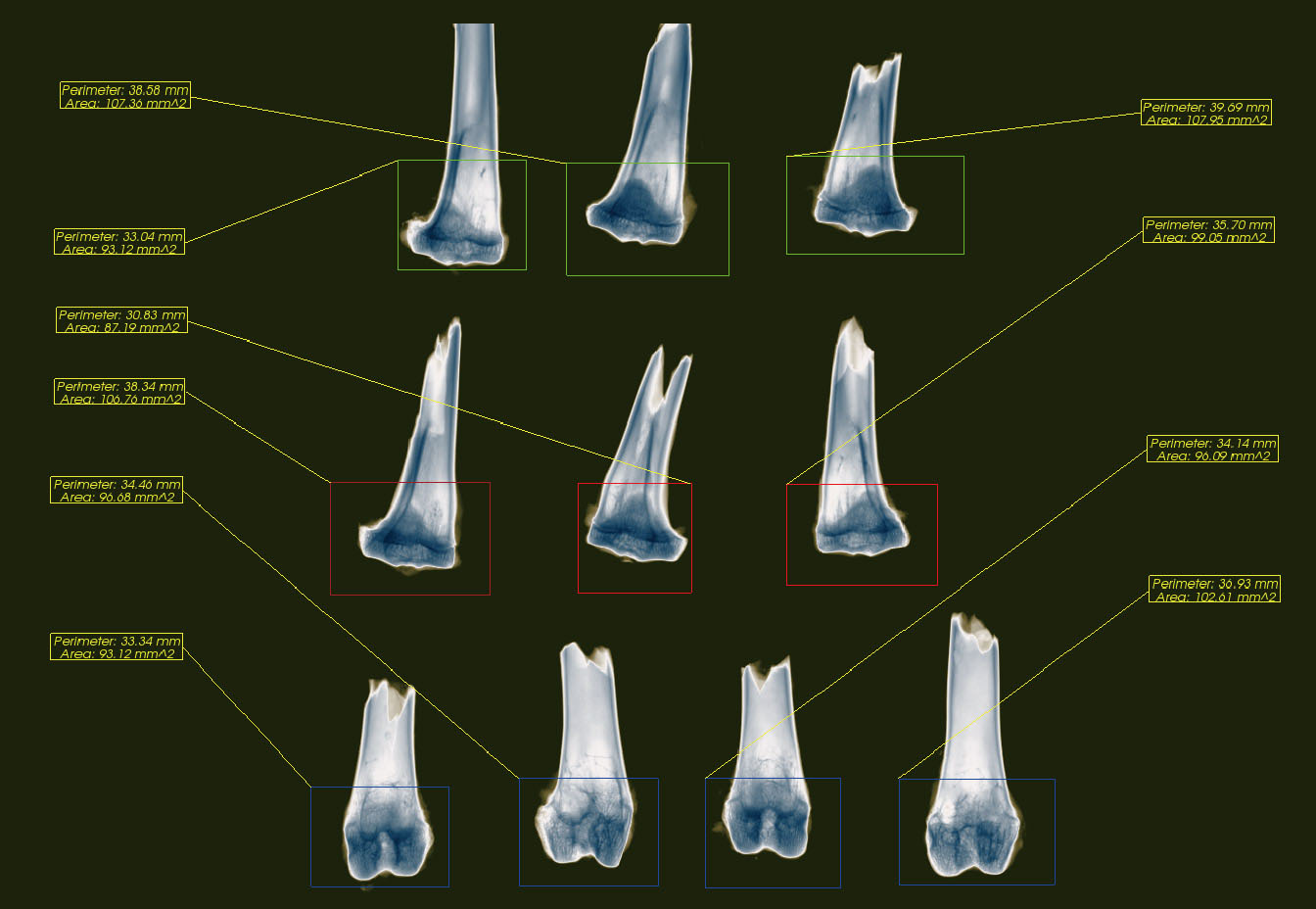
It features wide-field imaging and supports the simultaneous selection of multiple ROIs (regions of interest) on the image for the calculation of bone density or body composition parameters, which is used for related quantitative research.
The operations performed on the image (such as zooming in, zooming out, rotating, etc.), the delineation of the region of interest, and the measured data (such as perimeter, area, arbitrary angle, etc.) can be saved along with the image, ensuring data traceability.
100% self-shielding safety protection. The entire machine is designed with lead protection to ensure the safety of experiments and personnel, making it suitable for ordinary laboratories.

 中文
中文


